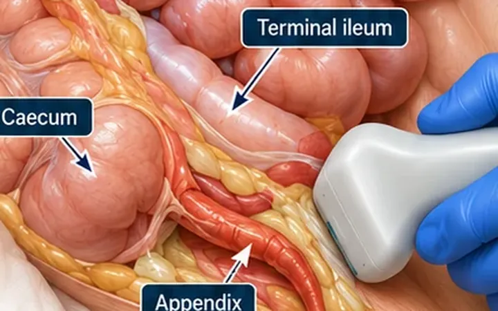
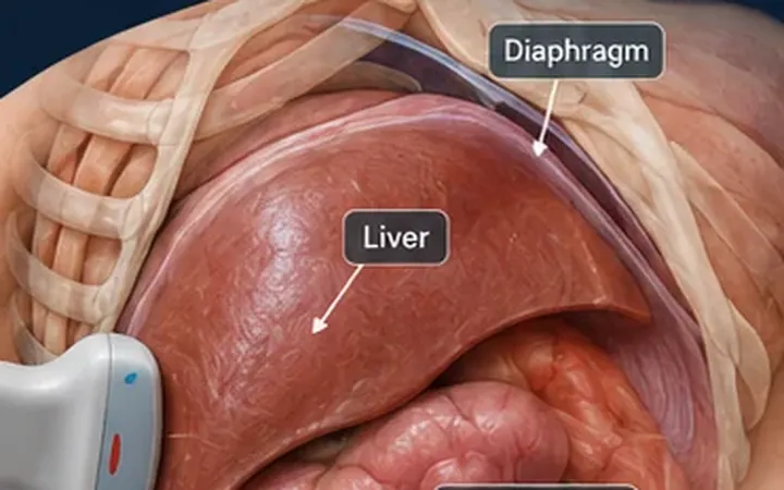
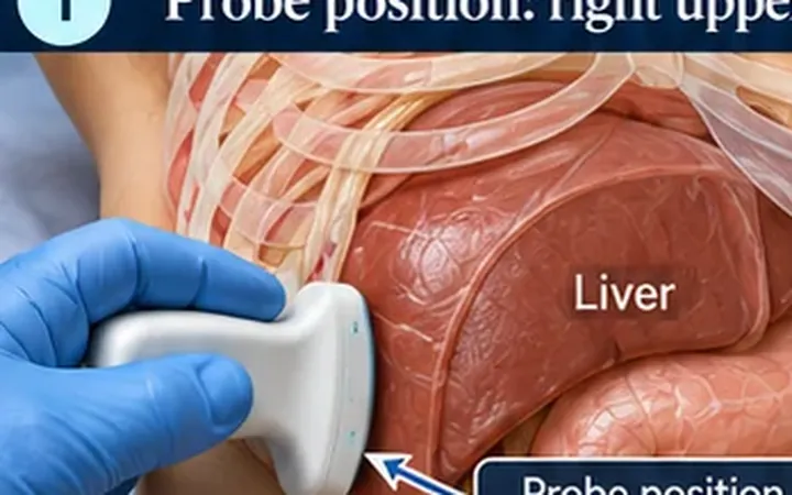
Intussusception is the commonest cause of bowel obstruction in children between about three months and three years. One segment of bowel telescopes into the next, usually the ileum into the colon (ileocolic). Delay risks ischaemia and perforation. Emergency physician ultrasound performs well: a 2020 meta-analysis found a sensitivity of 94.9% and a specificity of 99.1% for ileocolic intussusception.
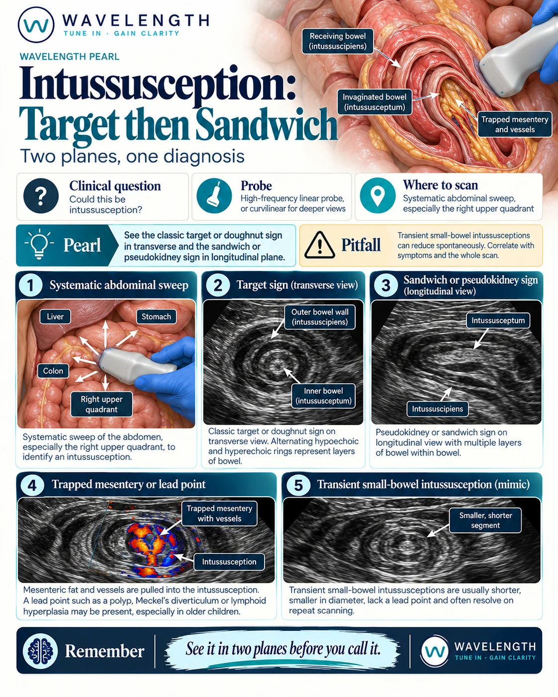
Scan images in the graphic are illustrations, not patient scans.
The scan
- Use the high-frequency linear probe. Switch to curvilinear for a larger child.
- Start in the right lower quadrant and follow the colon up to the right upper quadrant, across the transverse colon and down the left side. Most ileocolic intussusceptions sit on the right, under the liver.
- Sweep in transverse first, then turn 90° over anything you find.
- Use gentle, steady pressure. A distressed child tolerates the scan better on a parent's lap.
Reading the view
- Target or doughnut sign. In transverse, rings of alternating bright and dark layers: bowel inside bowel. Ileocolic intussusception usually measures 2.5 cm or more across.
- Sandwich or pseudokidney sign. In long axis, layers of bowel lying inside each other.
- Trapped mesentery. Bright fat and vessels pulled into the centre. Colour Doppler shows flow in the trapped vessels.
- Lead point. A mass, polyp, Meckel's diverticulum or large lymph nodes. More likely outside the usual age range.
Acting on the result
A positive scan in an unwell child needs urgent paediatric surgical and radiology referral for reduction. A negative bedside scan in a child with a convincing story still needs formal imaging.