Unrecognised oesophageal intubation kills. Waveform capnography is the standard for confirming tube position, and UK and international airway guidance is clear: no trace means wrong place. Capnography has limits in cardiac arrest, when low flow gives a flat trace even with the tube in the trachea. Ultrasound of the neck adds a quick, direct look.
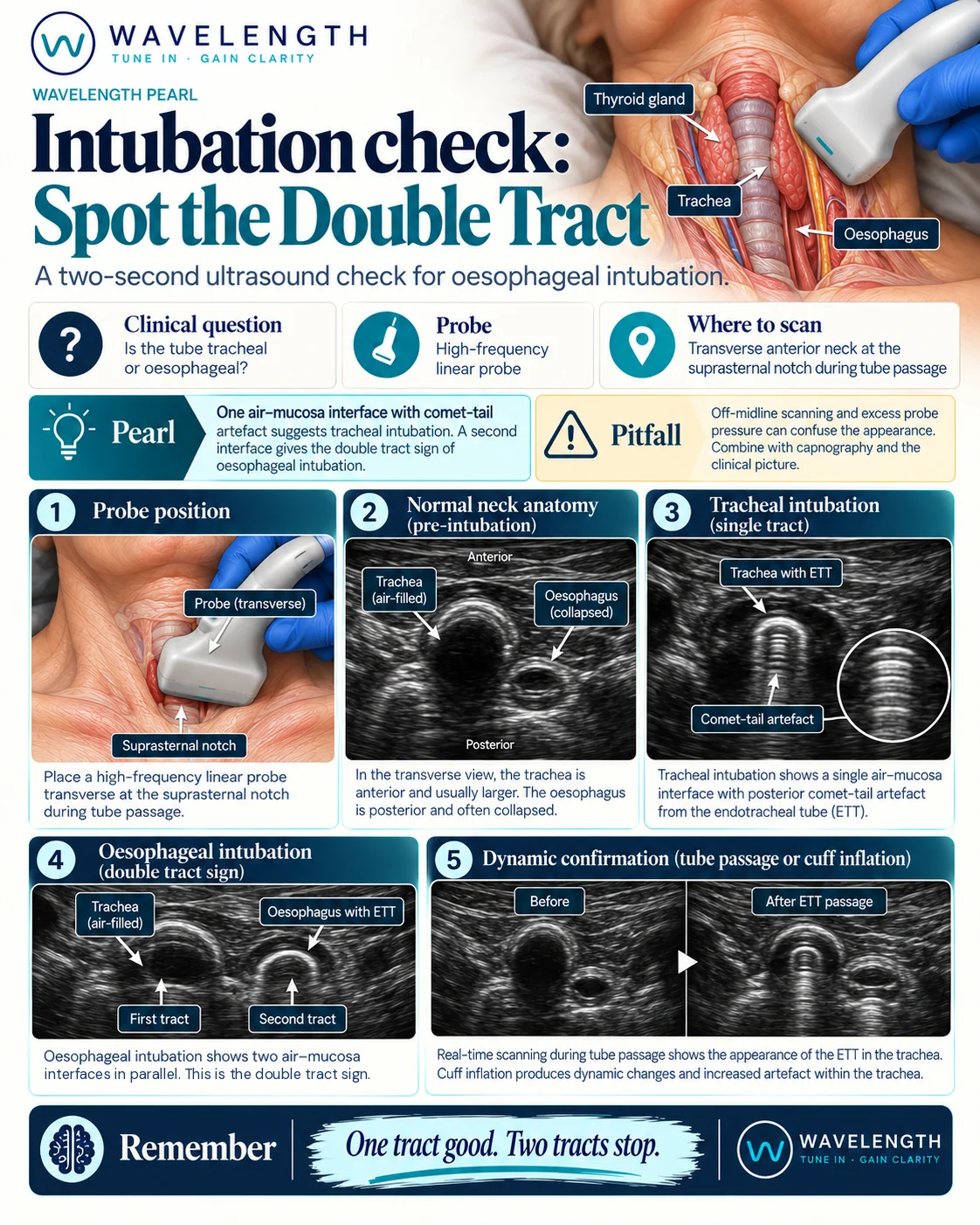
Scan images in the graphic are illustrations, not patient scans.
The scan
- Use the linear probe, transverse, just above the suprasternal notch.
- Before intubation, find the trachea, a bright curved line with reverberation behind it, in the midline.
- Find the oesophagus, usually behind and to the left of the trachea: a round structure with layered walls.
- Hold the probe still during intubation and watch as the tube passes.
- Confirm with waveform capnography.
Reading the view
- Tracheal intubation. One air-mucosa interface in the midline. The tube passing makes a brief flutter in the trachea. The oesophagus stays collapsed.
- Oesophageal intubation. A second bright curved line with reverberation appears beside the trachea, where the oesophagus was. Two air shadows sit side by side. This is the double tract sign.
After the tube is in
Scan both sides of the chest. Lung sliding on both sides shows both lungs are ventilated. Sliding on the right only, with a lung pulse on the left, suggests the tube sits in the right main bronchus.