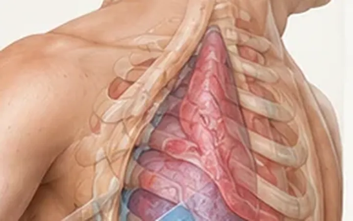
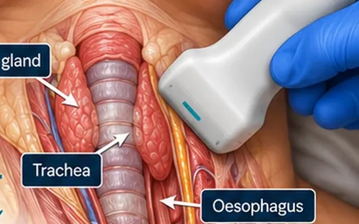
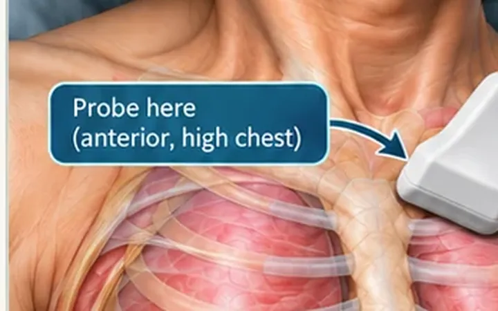
The diaphragm does most of the work of breathing. Phrenic nerve injury, an interscalene block, cardiac or thoracic surgery, neuromuscular disease and prolonged ventilation all weaken it. A chest X-ray shows a raised hemidiaphragm but not how it moves. Ultrasound shows movement directly.
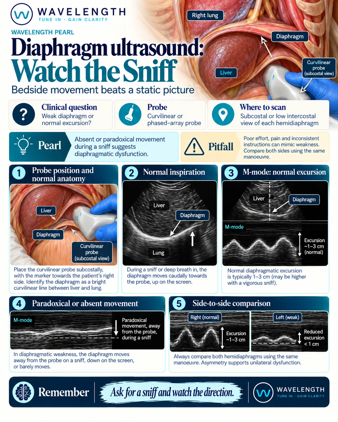
Scan images in the graphic are illustrations, not patient scans.
The scan
- Use the curvilinear or phased-array probe.
- Right side: place the probe below the right costal margin, between the mid-clavicular and anterior axillary lines, and aim up and back towards the dome. Use the liver as a window.
- Left side: use the spleen as a window, from a lower intercostal or subcostal position.
- Find the diaphragm, the bright curved line deep to the liver or spleen.
- Place the M-mode line through the dome.
- Record quiet breathing, a sharp sniff, then a deep breath. Repeat on the other side.
Which way is normal
The probe sits below the diaphragm. On inspiration the diaphragm contracts and moves towards the abdomen, which is towards the probe and up on the screen. The M-mode trace rises with each breath in.
Reading the view
- Normal: clear movement towards the probe on inspiration, similar on both sides.
- Weak: small movement, less than the other side.
- Paralysed: no movement on quiet breathing, and on a sniff the diaphragm moves away from the probe, down on the screen. This is paradoxical movement.