You have found the bat, watched the pleural line, and seen no sliding. Many clinicians stop here and call a pneumothorax. Absent sliding tells you something is wrong. It does not tell you what.
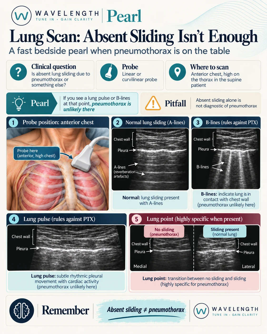
Scan images in the graphic are illustrations, not patient scans.
Where to look
In a supine patient, free air rises to the front of the chest. Start with the linear or curvilinear probe in the second to fourth intercostal spaces in the mid-clavicular line, probe head to foot across two ribs. Scan both sides.
Four signs, two questions
Each sign answers one question: are the two layers of pleura touching under the probe?
- Lung sliding. Shimmer along the pleural line as the lung moves with breathing. The layers touch.
- B-lines. Bright vertical lines from the pleural line to the bottom of the screen, moving with breathing. They start at the lung surface, so the lung is against the chest wall. The layers touch.
- Lung pulse. A subtle flicker of the pleural line in time with the heartbeat, with no sliding. The heart moves the lung, so the lung is against the chest wall. The layers touch.
- Lung point. The place where absent sliding meets sliding, as the lung edge moves in and out of view with breathing. This confirms pneumothorax.
When sliding is absent without a pneumothorax
- The patient is not breathing, or is holding their breath.
- Right main bronchus intubation. The left lung does not ventilate, so it shows no sliding but a lung pulse.
- Pleural adhesions after infection, surgery or pleurodesis.
- Severe consolidation, contusion or large bullae.
Use M-mode
Place the M-mode line across the pleural line. A moving lung gives a grainy pattern below the line, the seashore sign. No movement gives flat horizontal lines all the way down, the barcode sign. At the lung point, the trace switches between the two with breathing.