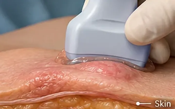
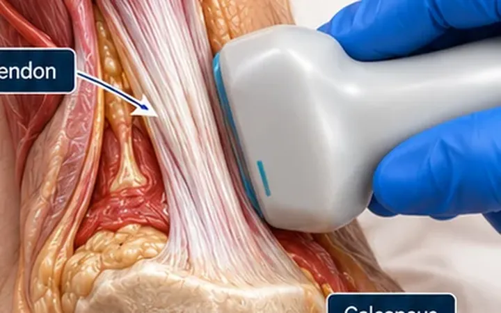
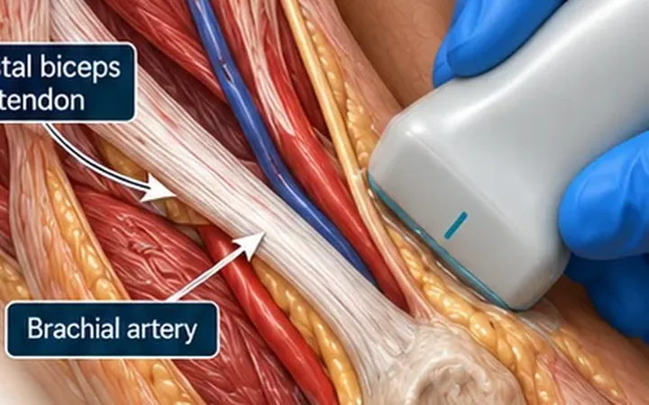
Examination of the hip is difficult, especially in a crying child. Ultrasound shows fluid in the joint in minutes and guides aspiration. It does not tell you what the fluid is.
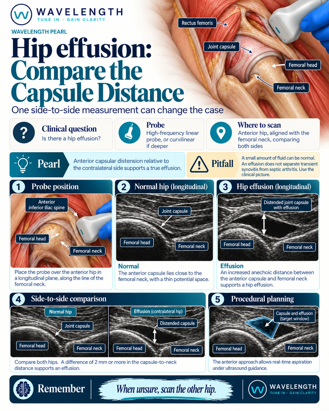
Scan images in the graphic are illustrations, not patient scans.
The scan
- Lie the patient supine with the hip in neutral, or slightly flexed and externally rotated.
- Use the linear probe in children and slim adults, the curvilinear probe in larger patients.
- Place the probe just below the inguinal ligament, lateral to the femoral vessels, along the long axis of the femoral neck, marker to the head.
- Find the femoral head, a bright curve, and the femoral neck running from it. The anterior capsule lies over the neck.
- Measure from the front of the femoral neck to the outer edge of the capsule.
- Scan the other hip with the same settings.
Reading the view
- Normal: the capsule lies close to the neck and follows its concave curve.
- Effusion: a dark space lifts the capsule off the neck, and the capsule bulges outwards.
- In children, ACEP guidance defines an effusion as a capsule-to-neck distance over 5 mm, or more than 2 mm wider than the other side.
Guided aspiration
The anterior recess is the target. Find the femoral vessels with colour Doppler, medial to the joint, and use an in-plane approach from lateral to medial. Follow your local orthopaedic pathway.