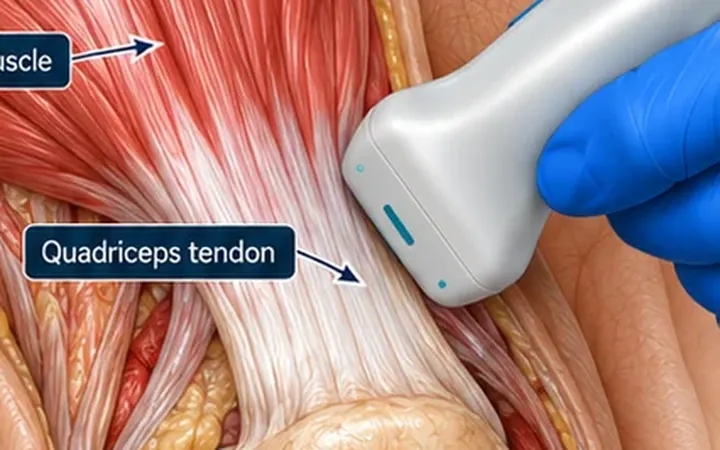
A hot, swollen knee needs an answer to two questions: is there fluid, and where is the safest place to aspirate it? The suprapatellar recess answers both. It connects with the knee joint and fills first when fluid builds up.
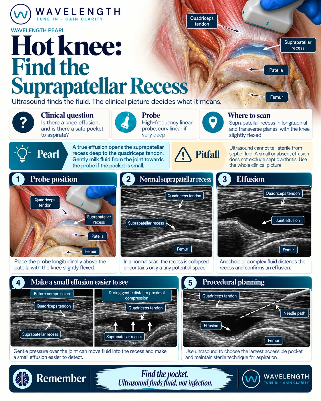
Scan images in the graphic are illustrations, not patient scans.
The scan
- Lie the patient supine with the knee flexed about 20 to 30° over a pillow. Slight flexion pushes fluid up into the recess.
- Place the linear probe in long axis just above the patella, over the quadriceps tendon.
- Find the bright, fibrillar quadriceps tendon, the top of the patella and the femur deep to them.
- Look between the tendon and the femur, under the fat pads. This is the suprapatellar recess.
- Turn to transverse and sweep medially and laterally to find the largest pocket.
- If the pocket is small, press gently on either side of the patella to squeeze fluid up into the recess.
Reading the view
- Normal: the recess is collapsed, a thin dark line between the fat pads.
- Effusion: a black or grey space opens deep to the tendon and spreads out under it. It compresses with the probe and moves with pressure.
- Complex fluid: debris, strands or a fluid level. Infection, blood and crystals all cause this.
- Synovial thickening: grey tissue that does not compress or move with pressure.
Guided aspiration
Use the largest accessible pocket, usually superolateral. Use full aseptic technique, keep the needle in plane, and watch the tip enter the fluid. Send fluid for microscopy, Gram stain, culture and crystals.