Tendons are bright and fibrillar on ultrasound only when the beam meets them at 90°. Tilt the probe a few degrees and a healthy tendon turns dark. This is anisotropy, the most common trap in musculoskeletal ultrasound.
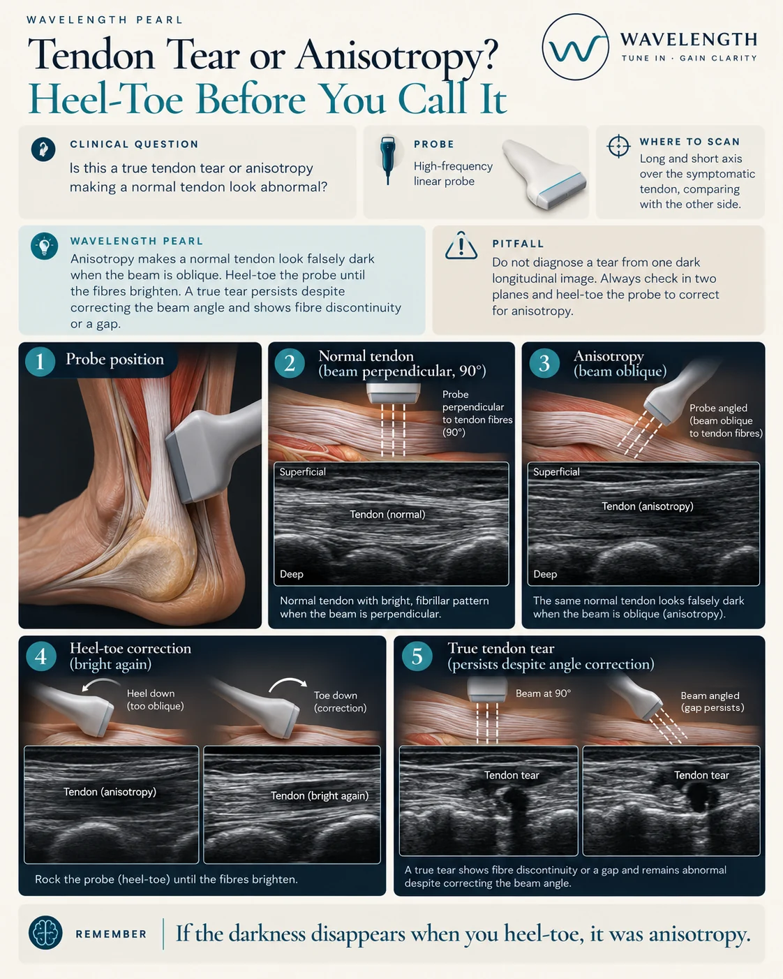
Scan images in the graphic are illustrations, not patient scans.
The scan
- Use the high-frequency linear probe with plenty of gel.
- Scan the tendon in long axis, then turn 90° to short axis.
- Where the tendon curves, rock the probe along its length: press the heel of the probe, then the toe, until the fibres turn bright.
- In short axis, tilt the probe from side to side for the same reason.
- Compare with the other side.
- Move the joint gently and watch the tendon glide.
Reading the view
- Normal. Bright, parallel fibres in long axis, and a bright speckled oval in short axis.
- Anisotropy. The tendon turns dark when the beam is oblique, and bright again when you correct the angle.
- Tear. Fibres break, leaving a dark gap or a defect which stays whatever the angle. In a full tear the ends separate, and movement of the joint shows they do not glide together.
Use anisotropy to your advantage
A nerve and a tendon look alike in short axis. Tilt the probe. The tendon darkens with a small change in angle, while the nerve changes little. This helps when you scan the wrist or before a nerve block.