Small bowel obstruction is fluid-filled. Fluid carries ultrasound well, so the dilated loops show up clearly, often before an X-ray shows the classic pattern. The movement inside the bowel helps separate mechanical obstruction from ileus.
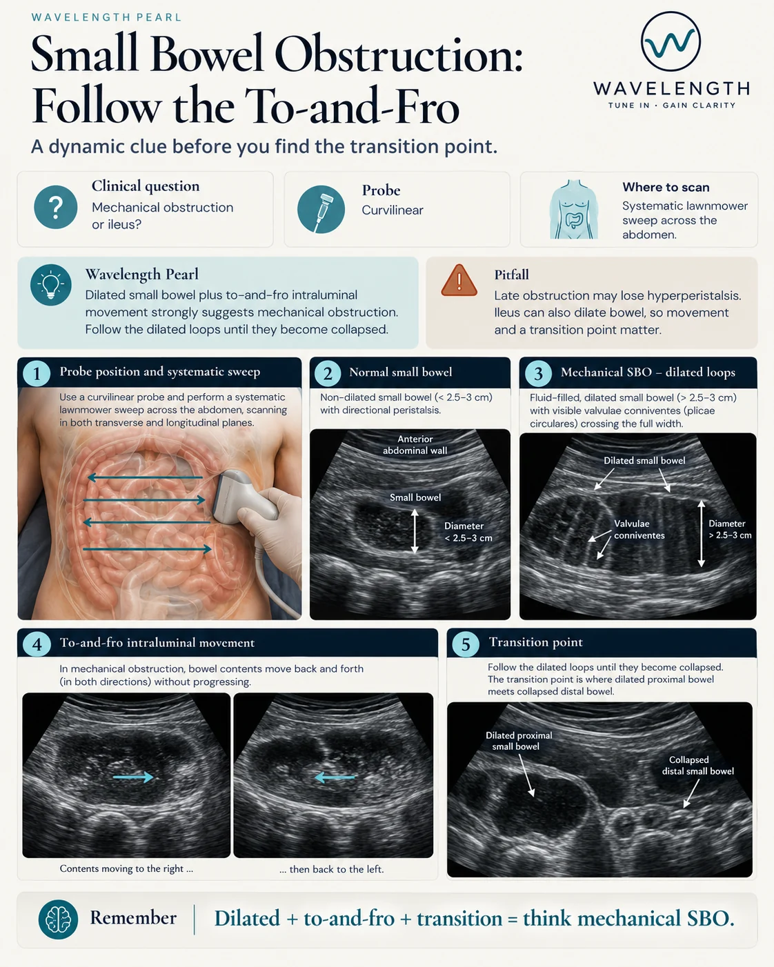
Scan images in the graphic are illustrations, not patient scans.
The scan
- Use the curvilinear probe.
- Mow the lawn: sweep the whole abdomen in rows, top to bottom, in transverse and then in longitudinal.
- Measure the widest small bowel loop from outer wall to outer wall.
- Watch the contents of a dilated loop for 10 to 20 seconds.
- Follow the dilated loops until they become collapsed.
What to look for
- Dilated loops. Small bowel over 2.5 cm across is dilated. Fluid-filled loops with the valvulae conniventes, thin folds crossing the full width, look like a keyboard or a ladder.
- To-and-fro movement. The bowel contents move back and forth without moving on. Strong, ineffective peristalsis against a blockage causes this.
- Transition point. Dilated bowel meets collapsed bowel. The cause often sits here.
- Free fluid between loops. Fluid between dilated loops, alongside a thick bowel wall or absent peristalsis, raises concern for strangulation.
Ileus or obstruction?
In ileus, the bowel dilates without a blockage. Peristalsis is reduced or absent all along the bowel, with no transition point, and both small and large bowel often dilate.