A chest X-ray in a supine patient misses a moderate pleural effusion, and it does not separate fluid from collapsed lung. Ultrasound answers both questions at the bedside, often in under a minute.
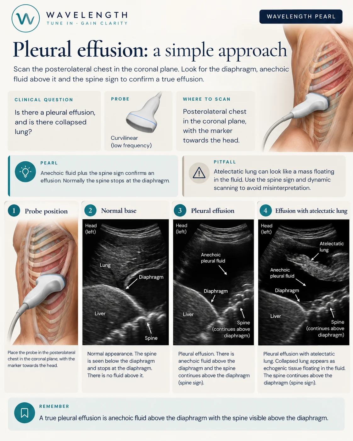
Scan images in the graphic are illustrations, not patient scans.
The scan
- Use the curvilinear probe, or the phased array between narrow ribs.
- Place it in the posterior axillary line, low on the chest, in the coronal plane with the marker to the head. In a supine patient, push the probe as far back as the bed allows. Fluid collects posteriorly.
- Find the diaphragm first: the bright curved line above the liver on the right, or the spleen on the left.
- Look above the diaphragm.
Reading the view
- Normal. Above the diaphragm you see air-filled lung, which moves with breathing (the curtain sign). The spine shows below the diaphragm and stops at it, because air blocks the beam.
- Effusion. A black, anechoic space sits above the diaphragm. The spine carries on above the diaphragm under the fluid. This is the spine sign.
- Effusion with collapsed lung. A wedge of tissue-like lung floats or flaps in the fluid.
- Consolidation. The lung itself looks like liver tissue, with bright air bronchograms. The spine sign appears here too, because consolidated lung also carries the beam. Look for the black fluid.
Beyond the diagnosis
- Fluid with internal strands or septations, or echoes which swirl, suggests a complex effusion, such as empyema or haemothorax in the right setting.
- Ultrasound guidance for pleural procedures is standard practice in the UK. Mark the site with the patient in the position you will use, and check for vessels with colour Doppler.