Hydronephrosis is dilatation of the kidney's collecting system, usually from obstruction further down. In renal colic it supports the diagnosis of an obstructing stone. In a febrile patient it raises concern for an infected, obstructed kidney. The skill is telling it apart from the black spaces which mimic it.
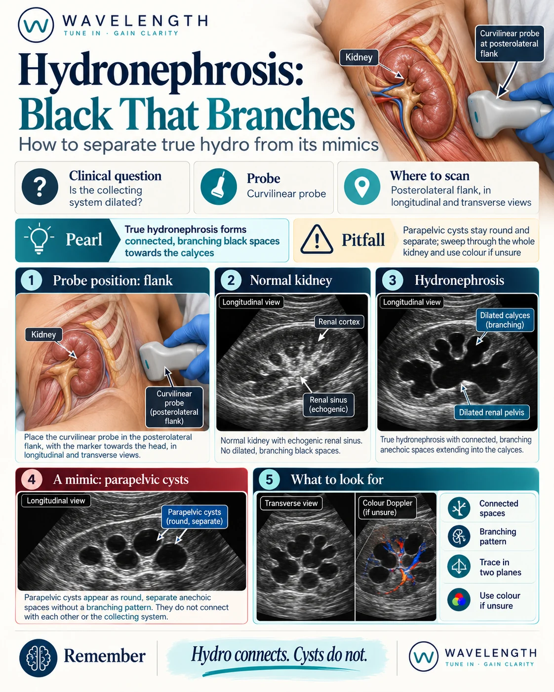
Scan images in the graphic are illustrations, not patient scans.
The scan
- Use the curvilinear probe with the patient supine.
- Right kidney: mid to posterior axillary line, using the liver as a window, marker to the head.
- Left kidney: sits higher and further back. Start in the posterior axillary line, higher on the flank, and use the spleen as a window. Put your knuckles on the bed.
- Sweep through each kidney in long axis, then turn 90° and sweep in short axis.
- Compare the two sides.
- Scan the bladder. A full bladder causes mild dilatation on both sides. Rescan after the patient has voided.
Reading the kidney
- Normal. A grey cortex around a bright, echogenic centre, the renal sinus. No black spaces inside the sinus.
- Mild. The renal pelvis is black and the calyces start to fill.
- Moderate. Black, rounded calyces join the pelvis, like a bunch of flowers on their stems.
- Severe. Large black spaces with a thin rim of cortex.
Mimics
- Parapelvic cysts. Round, separate, and not connected to each other or the pelvis.
- Prominent renal sinus vessels. Colour Doppler fills vessels with colour. A dilated collecting system stays black.
- Extrarenal pelvis. A renal pelvis lying outside the kidney looks dilated, while the calyces stay normal.
The older patient with renal colic
A patient over 50 with first-time renal colic needs their aorta scanned. A leaking abdominal aortic aneurysm presents with flank pain and blood in the urine. Thirty seconds on the aorta prevents a fatal miss.